Friday, November 12, 2021

Antibiotic Resistant Genes in Your Water!?

By Ruby Ayala
The experimental design of three transformation model systems designed to assess the effects of chlorine-based disinfectants on the spread of antibiotic-resistant genes. The figure is taken from Zhang et al. (2021)

Antibiotic-resistant bacteria are a rising threat to public health worldwide. Chlorination is a method of disinfection that has been widely used to inactivate bacteria and pathogens in wastewater treatment plants; however, the disinfection process can damage antibiotic-resistant bacteria and result in the release of antibiotic-resistant genes into the water that would be readily available for uptake by other bacteria through their cellular membrane. In a study by Zhang and colleagues (2021), they used plasmid-encoded antibiotic-resistant genes and a recipient bacteria in three different transformation systems to determine whether chlorination influenced the spread of antibiotic-resistant genes in conditions mimicking drinking water and the effluent of wastewater treatments. After verifying the recipient bacteria took up the plasmid and showed signs of resistance to two antibiotics, ampicillin and tetracycline, the results revealed that at relevant concentrations, chlorine-based disinfection can promote the natural transformation of antibiotic-resistant genes among bacteria found in water. This could be attributed to the fact that disinfection induces a series of cell responses and bacterial membrane damage that enhance the bacteria's uptake of antibiotic-resistant genes. The findings of this study give scientists a better understanding of the role of disinfectants and their relation to the dispersal of antibiotic resistance in bacteria. With the knowledge gained, better management of disinfection practices in water systems can be strategized and applied to protect the health of humans and future generations.

Original article: 

Zhang S, Wang Y, Lu J, Yu Z, Song H, Bond PL, Guo J. 2021. Chlorine disinfection facilitates natural transformation through ROS-mediated oxidative stress. The ISME Journal 15:2969-2985. 


Saving the Honey Bees with Bacteria

 By Ruby Ayala


A honey bee with a Varroa mite. Photo taken by Alex Wild from The University of Texas at Austin

Honey bees are very critical pollinators and play a huge role in global food production. Without honey bees, dozens of agricultural crops ranging from fruits to nuts to vegetables would either decrease substantially or vanish all together. Unfortunately, our friendly neighborhood pollinators are experiencing wide population declines due to parasites, such as Varroa mites, and pathogens, such as the RNA virus that causes wing deformity. The increasing loss of the honey bees has become a problem for agriculture; therefore, Leonard and colleagues from the University of Texas at Austin conducted a study wherein they evaluated the honey bee's survival rate after having engineered bacteria that inhabit their gut microbiome to combat varroa mite infestations and wing deformity. The team engineered two different bacterial strains for the honey bees to ingest; one that targets the RNA virus and one that targets the Varroa mites. The engineered bacteria have a symbiotic relationship with the honeybee and produce double-stranded DNA that alters gene expression and triggers RNA interference immune responses. The results of the study showed that compared with the control bees, the bees with wing deformity treated with the engineered bacterial strain were 36.5% more likely to survive a longer period of time (10 days). Also, the Varroa mites on bees treated with the mite-targeting strain were 70% more likely to die compared to the mites feeding on the control bees. The findings of this research not only provide a possible solution to decrease the loss of honey bee populations but also gives other researchers an insight into how bacteria can be altered to save other species from disease.

Original article:

Leonard SP, Powell JE, Perutka J, Geng P, Heckmann LC, Horak RD, Davies BW, Ellington AD, Barrick JE, Moran NA. 2020. Engineered Symbionts Activate Honey Bee Immunity and Limit Pathogens. Science 367:573-576.


Deep-Sea Deep Sequencing: A Showcase of Extraordinary Viral Diversity

 By: Umberto Fasci

 
Phylogenetic analysis of viral contigs from marine sediments, marine water, freshwater and terrestrial soil - proteome-wide similarity relationships. Figure taken from Zheng et al. 2021.


 This study with the fascinating aim to improve the knowledge of the virosphere in deep-sea sediments, investigates the viral diversity at both gene and genomic levels in the deep-sea sediments of the Southwest Indian Ocean. This alone seems ambitious. However, with the decreasing cost and increasing data acquisition of sequencing technologies, deep sequencing has become valuable for this use case. From their deep sequencing analysis, the researchers here found a large number of unclassified viral groups with a total of 1106 viral contigs. Amazingly, 217 of these expressed complete viral genomes with none clustered with any known viral genome. It was also found that over two thirds of the ORFs within these viral contigs encode for no known functions. This by itself is extraordinary, suggesting the the deep-sea sediments represent an enormous site for novel viral genotypes. Ultimately, this study opens a box which requires the right tools to access. With this, future related studies should explore how the novel viral metabolic genes found here may be involved in energy production, or amino acid synthesis pathways at this level of the marine environment. Elucidating this may improve our understanding of how involved these novel viral genes are in the marine sediment environment.


Zheng, X., Liu, W., Dai, X., Zhu, Yaxin, Wang, J., Zhu, Y., et al. (2021) Extraordinary diversity of viruses in deep-sea sediments as revealed by metagenomics without prior virion separation. Environ Microbiol 23: 728–743.


Lacking Phosphorus? These Microbes Might be the Answer

 By: Umberto Fasci

 
The distribution and activity of isolated PSMicro strains in bamboo rhizosphere. A. Isolated PSmicro strains in bamboo rhizosphere. B. Relative abundances of isolated PSMicro strains in bamboo rhizosphere. C. Variation in phosphorus solubilization by Pseudomonas sp. in bamboo rhizosphere. D. Variation in phosphorus solubilization by Burkholderia sp. in bamboo rhizosphere. Figure taken from Xing et al. 2021.


To understand the importance of what this study investigates, I will illuminate the importance of phosphorus-solubilizing microorganisms (PSMicros). PSMicros are essential in assisting associated plants to resist phosphorus deficiency in soils. On the whole, this study investigates the microbial diversity of PSMicro strains from several bamboo rhizosphere sites. The study showed great variation in microbial diversity between these bamboo rhizosphere sites where 52 PSMicro strains were isolated and identified. Among these isolations, 10 bacteria genera and 4 fungal genera were identified. From this, the researchers of this study found that Bacillus, Kluyvera, Buttiauxella, Meyerozyma and Penicillium species were most readily utilized to supply plant-usable phosphorus from both organic and inorganic phosphorus sources. As such, this study identifies PSMicro strains which can be possibly used in ecosystem restoration. A good step for future research into this subject should take these results as reference, and to conduct a more thorough investigation with metagenomic techniques. The potential impacts alone this study has should inspire future research as such.


Xing, Y., Shi, W., Zhu, Y., Wang, F., Wu, H., and Ying, Y. (2021) Screening and activity assessing of phosphorus availability improving microorganisms associated with bamboo rhizosphere in subtropical China. Environ Microbiol 23: 6074–6088.

Saturday, November 6, 2021

Bacteria that Aid in Petroleum Polluted Soil

 By: Melissa Villareal 

The figure shows the residual Naphthalene (NAP) percentage after an 8 day incubation. By day 8, there was a significant reduction of the percentage of NAP compared to the sterile control group. Figure taken from Cai et al. 2021.


Environment pollution is an increasing problem with industrialization and globalization. The world relies on petroleum to operate machinery and vehicles. This can lead to petroleum contamination in soil, and it is a serious problem. Petroleum contamination contain organic pollutants such as polycyclic aromatic hydrocarbons (PAHs) that pose a serious threat to human health. PAHs are toxic and can cause cancer, DNA mutations and other serious effects on health.

A study by Cai and colleagues (2021) identified a bacteria species that can degrade a type of PAH called naphthalene (NAP). This uncultured bacteria is a Gamma-Proteobacterium species, and it is a key NAP degrader. This is a form of bioremediation mechanism that is found in the natural environment. Their metabolism is able to break down this organic pollutant.

The next step of this research is to identify more bio-degraders of PAH, and being able to culture these bacteria species. Overall, this research is very important to efficiently and safely clean up pollutants from the soil. This can prevent human health consequences, and protect the ecosystem.    


Original Article:

Cai, X., Li, J., Guan, F., Luo, X.and Yuan, Y. (2021) Unveiling metabolic characteristics of an uncultured Gammaproteobacterium responsible for in situ  PAH biodegradation in petroleum polluted soil. Environ Microbiol.

Soil Bacteria Can Fight Climate Change

 By: Melissa Villareal 

The figure shows the detected microbial communities from the three desert plant species. The red points represent the same microbes shared by the three desert plants. Figure taken from Sun et al. 2021.


Climate change has increased the desertification of arid regions. Desertification is the process by which fertile areas degrade and lose bio-productivity. The lands become infertile and hostile to plants and animals. This rapid increase of desertification poses an environmental threat. 

Desert plants can aid in recolonizing desert areas to combat the loss of biodiversity. Three desert plant species continue to thrive even under increased stress caused by climate change because they have high adaptability. A study by Sun and colleagues (2021) show that the microbiota living in these desert plants are key to their survival. These bacteria provide essential nutrients, aid in plant hormone production and protect the hosts from harmful pathogens. These bacteria species can sustain plant growth under extreme abiotic stress! So far two bacteria species have been identified: Streptomyces eurythermus and Streptomyces flaveus

Finding methods to combat desertification is important to maintain biodiversity in these arid areas, and offers possible solutions in desert management. Lastly, studying bacterial communities that aid in plant growth in extreme abiotic stress can help the agriculture industries. Microbial tools can increase agricultural productivity in a safer and more sustainable way.


Original Article:

Sun, X., Pei, J., Zhao, L., Ahmad, B.and Huang, L. (2021) Fighting climate change: soil bacteria communities and topography play a role in plant colonization of desert areas. Environ Microbiol.

Tuesday, October 26, 2021

Neovaginal Secretions Contain Diverse Communities of Anaerobic Bacteria

  By: Chelsea Alcala


Microbial community structures found via mass spectrometry in neovaginal, rectal, and vaginal compartments of transgender and cisgender women. Figure taken from Birse et al. 2020.


 Women who undergo gender reassignment surgery have fundamentally different vaginas from that of ciswomen, called neovaginas. Neovaginas are constructed (neovaginoplasty) through penile inversion, scrotal/sigmoid colon grafts, or a combination of the former mentioned. However, unlike ciswomen, not much is known about this organ's microbiome which plays a critical role in the sustainability of infection(s), inflammation, epithelial barrier function, & overall neovaginal health. Using a metaproteomics technique this study, carried out by Birse and colleagues, mapped and classified the microbial composition of neovaginal/rectal secretions (transwomen) and compared them with vaginal secretions (ciswomen). Metaproteomics identified 541 distinct bacterial proteins from 38 taxa. The most abundant taxa found in neovaginas included; Porphyromonas (30.2%), Peptostreptococcus (9.2%), Prevotella (9.0%), Mobiluncus (8.0%), and Jonquetella (7.2%). While cis-vaginal samples & rectal samples contained predominantly Lactobacillus, Gardnerella, Prevotella, and Roseburia (see figure above for specifications). Moreover, similar to characteristics found in uncircumcised penises and cis-vaginas (with bacterial vaginosis), penile skin-lined neovaginas were found to have diverse polymicrobial communities (primarily anaerobic), increased immune activation pathways, and decreased epithelial barrier function. Research like this is extremely important in the world of medicine due to the fact that transgender health care is often overlooked, understudied, and neglected. By not conducting research to determine the composition of the neovaginal microbiome or similar research, the scientific community would be marginalizing all trans-women and frankly ignoring a vast gap in human knowledge. Research much like this one could significantly alter how we treat inflammation/infection in the neovaginal environment, improving transgender health & wellness as a whole.


Original Article:

Birse, K. D., Kratzer, K., Zuend, C. F., Mutch, S., Noël-Romas, L., Lamont, A., et al. (2020). The neovaginal microbiome of transgender women post-gender reassignment surgery. Microbiome, 8: 1-13.


Monday, October 25, 2021

Climate Change Affecting Snow Covered Ecosystems

 By: Jacqueline Martinez

Graphs from the study that demonstrate the correlations of bacteria, fungi, soil carbon to nitrogen ratio, and soil pH, which are according to the snowmelt timing.


Due to climate change affecting the warmth of the planet, there are side effects to this, which are the earlier summer temperatures that cause snow to melt earlier. Since snow begins to melt at an earlier date than expected, this also creates change to the soil and its microbial communities because it is being exposed sooner especially in alpine areas. Moreover, the shift in temperatures at different dates allows the soil to respond to these changes accordingly with the circumstances, which may cause a disturbance at an ecological level. The study conducted by Broadbent and colleagues had used plots of land to demonstrate the effects of early snowmelts happening during the spring where there was three manipulation treatments: snow removal, snow addition, and untreated control treatment. The findings ended up showing the correlation of the change of when the spring snow melting happens and nitrogen cycling. Furthermore, the findings showed there could be future changes in both carbon and nitrogen in snow covered areas and will affect the natural order of things in an ecosystem, such as plant growth.


Journal Article:

Broadbent, A. A. D., Snell, H. S. K., Michas, A., Pritchard, W. J., Newbold, L., Cordero, I., Goodall, T., Shallhart, N., Kaufmann, R., Griffiths, R. I., Schloter, M., Bahn, M., & Bardgett, R. D. (2021). Climate change alters temporal dynamics of alpine soil microbial functioning and biogeochemical cycling via earlier snowmelt. The ISME Journal, 15, 2264–2275. https://doi.org/10.1038/s41396-021-00922-0.


The Effect of Nanoscale Hydroxyapatite in Infected Tomatoes

 By: Andrea Martinez

The figure shows the PAL overall activity (graphs A and B) and the phenolic content (graphs C and D) that is found in the tomato roots and shoots. This is the content found in both the healthy and the diseased groups that were used to find the data. Figure from Ma, et al., 2021. 


    This study was used to find the mechanisms in which large and small-sized nanoscale hydroxyapatite can suppress Fusarium-induced wilt disease in tomatoes. There were two separate groups that were used one had a low dose and another had a high dose which was distributed to tomatoes that were infected with this disease and the purpose was to see how the nanoscale hydroxyapatite would affect the shoot mass of the infected tomatoes. The exposure to both of the nanoscale hydroxyapatite sizes raised a significant amount of phenylalanine ammonia-lyase activity within the infected tomatoes. It was also found that the phenolic content increased and it was seen to increase the shoots in Fusarium-infected tomatoes by 30-80% and around 40-68%.  The large-size treatments remodeled the cell membrane in the infected tomatoes as a way to try and protect the tomatoes from the infection. 


References: 

https://pubs.acs.org/doi/pdf/10.1021/acs.est.1c00901

Zinc oxide nanoparticles ecotoxicological risk to aquatic fungi


By: Melissa Garcia


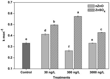
After 45-day exposure to nZnO and  ZnSO4 in three different concentrations, overall the rate of decomposition was faster when microcosms were exposed to ZnSO4. The decomposition rate was found the lowest in the microcosm with nZnO at 300 ng/ L. The highest decomposition rate was observed in microcosm with  ZnSO4Figure taken from Du et al. 2020


There is an ecotoxicological risk of zinc oxide nanoparticles (nZnO) in freshwater ecosystems. The toxicity from these nanoparticles can be attributed to release of ionic Zn, increase in oxidative stress, and its physiochemical properties. To understand the containments effect on aquatic fungi, researchers examined leaf litter decomposition, an important degradable nutrient in ecosystems. The study explored the rate of decomposition in leaf litter after exposed to nZnO and ZnSO4 in different environmental concentrations using a microcosm with artificial reconstituted fresh water (ARFW) along with a control microcosm. Scientists also explored the litters chemical components, changes of ionic Zn concentration, enzyme activity, fungal community and fungal biomass after exposure. Results demonstrated an increase of ionic Zn concentration over a 45 day exposure in ARFW with nZnO, and contrastingly a decrease of ionic concentration in ARFW with ZnSO4. The leaf litter rate of decomposition was decreased in microcosm with nZnO. Contrastingly, ZnSO4 demonstrated an increased decomposition rate. After exposure to concentration, an increase in carbon and nitrogen was observed in both contaminated microcosms. The fungal biomass increased in microcosm nZnO and decreased in microcosm with ZnSO4. After 45-day exposure, fungal diversity was enhanced when exposed to microcosm with nZnO and ZnSOcompared to the control microcosm. Researchers concluded even at the lowest concentration’s exposure to nZnO can impact leaf litter decomposition. These findings are important to highlight potential environmental toxicity risks from zinc oxide nanoparticles exposure.


Reference:

Du, J., Zhang, Y., Yin, Y., Zhang, J., Ma, H., Li, K., & Wan, N. (2020). Do environmental concentrations of zinc oxide nanoparticle pose ecotoxicological risk to aquatic fungi associated with leaf litter decomposition? Water Research178, 1–8. https://doi.org/10.1016/j.watres.2020.115840 


What Does Sunlight do to Antibiotics in the Environment?

By: Samantha Johnson

The figure above shows the the loss of antibiotic, oxytetracycline and streptomycin, potency in the presence of sunlight. Figure was taken from Khan et al. 2021.

Scientists predict that by 2050 antibiotic resistant-related deaths will be the number one killer in the world. A study by Khan and colleagues researched the effect that sunlight and UV radiation have on antibiotics commonly used to prevent the spread of citrus greening disease, a disease that affects citrus plants that is caused by a bacterium. The antibiotics tested were oxytetracycline and streptomycin. To test the antibiotics efficacy, they were applied to bacteria and left in sunlight or in darkness for 7 or 14 days. The figure above shows the results of this test. In the dark, oxytetracycline continues to have a high efficacy, but in sunlight its efficacy quickly decreases. Streptomycin only showed moderate efficacy loss in the sunlight and remained potent in darkness. Oxytetracycline was further studied with a UV light which found that the treated discs had a dramatic decrease in efficacy. These results highlight how long antibiotics may last outdoors and what may or may not leach into the ecosystem. The next steps in this research is to test more antibiotics and to relay pertinent information to appropriate agencies and consumers. Hopefully for all of humankind, a solution can be found to treat antibiotic resistance. 

Original Article:

Khan S. J., Osborn A. M., and Eswara P. J. (2021). Effect of Sunlight on the Efficacy of Commercial Antibiotics Used in Agriculture. Front. Microbiol. 12:645175.

Lighting The Way to Minimizing the Future Spread of Covid

 by: Ruby Ayala

Disinfection through the traditional use of chemicals (pictured on the left) vs disinfection through the use of UV light (pictured on the right).    Photo Credit: Jennifer M. Mccan/Penn State

With the ongoing cases of covid-19 in today's society, scientists are looking for ways to minimize the spread of the virus. One proposed method of disinfection is through the use of ultraviolet (UV) light radiation, which has been proven to be an effective tool in killing viruses. In fact, UV light technology has already been adopted to use for disinfection purposes in healthcare settings. however, due to the safety testing requirements of SARS-CoV-2 being limited to biosafety level 3 laboratories, data on the efficiency of UV light on covid disinfection is still limited. In a study by Ben Ma et al., they researched the effects of different far ultraviolet-C light wavelengths on the SARS-CoV-2 virus, as well as identified the murine hepatitis virus, which falls under a lower biosafety level, as a reliable substitution for covid-19 in further UV disinfection tests. For this study, virus samples were diluted in thin-film liquid and placed under the following five UV sources: a filtered KrCl excimer UV light, an unfiltered KrCl excimer light, a mercury UV lamp, and two UV LED systems. The results showed that all the UV lights were capable of inactivating the virus in the aqueous solution; however, both the krypton chlorine (KrCl) excimer UV lights produced the highest disinfection rates. Compared to the other far ultraviolet-C devices, the krypton chloride excimer lamp is fueled by molecules moving between different states of energy, so a UV wavelength at 222 nanometers is able to inflict greater viral protein and nucleic acid damage to the virus due to the wavelength's high energy. This finding is particularly important because this ultraviolet-C device is relatively safe for human exposure, thus it can be used in occupied public spaces, such as subways and elevators, to disinfect viruses in airborne droplets and contaminated surfaces. 

Original Article:
Ma, B., Gundy, P.M., Gerba, C. P., Sobsey, M. D., & Linden, K. G. (2021). UV Inactivation of SARS-CoV-2 across the UVC spectrum: KrCl* excimer, mercury-vapor, and LED sources. Applied and environmental microbiology, AEM0153221. Advance online publication. https://doi.org/10.1128/AEM.01532-21

Soil Methanogenesis Caused by Deforestation of the Amazon Rainforest for Agriculture

 By: Jaime Vargas

The figure has four graphs that demonstrate the relative abundances of methanogens in incubated soil samples from Pará and Rondônia. The y-axis is the abundance of methanogens, while the x-axis is the land type from which the soil was taken from. The PF stands for primary rainforest in green color, P stands for pasture in orange, and SF stands for secondary rainforest in blue. The bars with different colors within the graph demonstrate which genus of methanogen was more abundant in the soil samples. The two top graphs (A and B) had carbon dioxide (CO2) as a carbon source in the soil samples. The two bottom graphs (C and D) had sodium acetate (NaAOc) as the carbon source in the soil samples. Graphs A and C had soil samples from Pará, and graphs B and D had soil samples from Rondônia. Figure taken from Kroeger et. al., 2020.


The Amazon rainforest is one of the most diverse ecosystems in the world, but unfortunately this ecosystem has been reduced over time due to deforestation. The Amazon rainforest is known as a carbon sink, which is an area that takes up carbon compounds from the atmosphere like CO2. Deforestation in the Amazon rainforest leads to an increase in carbon compounds into the atmosphere by releasing the stored carbon, making the area a carbon source instead of a carbon sink. From deforestation new pastures and agricultural fields are born, and in the study by Kroeger et. al., the levels of methanogenesis are measured by comparing new pastures and the rainforest areas. The amount of methane produced can be linked to microbes in the soil, so the study focuses on culturing bacteria in different soil samples to determine if pasture soil increases methanogenesis or not. Soil samples were gathered from one cattle pasture and two rainforest regions called Pará and Rondônia. The samples were treated with distinct carbon sources and incubated; then DNA analyses were done to determine the abundance of methanogenic microbes in each soil sample. The results demonstrated that in pasture soil there was a higher abundance of methanogens compared to rainforest soils. The results show that loss of rainforest and increase in pastures leads to an increase in methanogenesis causing more green house gases to enter the atmosphere. Future research should focus on why pastures are more suitable for methanogens.

Reference:

Kroeger, M. E., Meredith, L. K., Meyer, K. M., Webster, K. D., de Camargo, P. B., de Souza, L. F., Tsai, S. M., van Haren, J., Saleska, S., Bohannan, B. J. M., Mazza Rodrigues, J. L., Berenguer, E., Barlow, J., Nüsslein Klaus, & Los Alamos National Lab. (LANL), Los Alamos, NM (United States). (2020). Rainforest-to-pasture conversion stimulates soil methanogenesis across the Brazilian Amazon. The ISME Journal, 15(3).

Tea Time: How the temperature of your tea affects biofilm colonization

 By: Angelica Leos

 Graphs A and B show the effect on glucosyltransferase (GTP) production by S. mutans and CFU counts respectively, after treatment with different steeping conditions of green and black tea. Figure taken from Kim et al. 2020.

Oral biofilms are responsible for infectious dental diseases such as periodontitis and peri-implantitis that lead to the loss of teeth. The primary bacterial pathogens that cause these disease along with others are Streptococcus mutans and Streptococcus sobrinus (Kim et al., 2020). A study performed by Kim and colleagues focused on the effects of tea extracts on oral biofilm colonization depending on the difference in steeping temperature. The researchers use two types of tea extract: green tea and black tea. The tea extracts were each prepared with a hot steeping and a cold steeping for a total of four treatments total. The researchers found that while both tea extracts decreased the total biofilm biomass of both strains, both tea extracts showed higher inhibition of the pathogens when prepared with hot steeping. This means that tea extracts prepared with hot steepings were more effective in the prevention of the establishment of Streptococcus mutans and Streptococcus sobrinus. This data emphasizes the potential role of natural foods in the prevention of pathogen-causing diseases for dental cavities. It is important to consider how the preparation of food and drinks can play a larger role in the microbial environments of the human body in order to combat disease and take care of overall human health.


Original Article:

 Mi-Ah Kim, Jae-Hwan Kim & Ok Hyung Nam (2020) Tea extracts differentially inhibit Streptococcus mutans and Streptococcus sobrinus biofilm colonization depending on the steeping temperature, Biofouling, 36:3, 256-265

A possible end to the pandemic?

 By: Alejandra Pena

a-c, inhibition of entry of lentiviral PVS in 293-T-ACE2 (a), Caco-2 (b) or Calu03 (c) cells by the serine protease inhibitor, camostat (green bars), or the cathepsin inhibitor, E64-d (purple bars). All assays were performed three times and are plotted as mean +s.d. The data that is shown are representative replicate (n=3). Data was normalized to no drug control (black bars). The statistics were shown by two-way ANOVA with various comparisons. d, Replication kinetics of SARS-CoV-2 WT in HAE cells. The cells were pretreated with either control media or media containing camostat for 1 h that was then infected at MOI of 0.1. (e) Caco-2 (f) Calu-3 (g) or HAES (h). Gene expression by RT-qPCR and normalized to B-actin. Figure taken from Peacock et al. 2021. 


SARS-CoV-2 entered the human population and basically consumed humanity as we know it. Coronaviruses are able to enter cells through their spike glycoprotein. In order for SARS-CoV-2 to enter the host, it requires a cleavage of the spike glycoprotein in the S1/S2 and S2’ cleave sites in order for membrane fusion to begin. Throughout the study, there was a combination of lentiviral pseudotypes with spike cleavage sites mutations and Vero-passaged SARS-CoV-2 virus variants used in order to further understand the molecular mechanism of polybasic cleavage sites of SARS-CoV-2 and their entry into the lungs. The study shows how SARS-CoV-2 viruses that lack the S1/S2 furin cleavage site showed lower titres from infected ferrets and were not transmitted onto other animals. The study then shows that TMPRSS2 is a useful drug target and although it would not prevent infection through the endosome, the pathway is crucial in preventing the replication of this virus in the airway. This study is crucial to society as the pandemic is still happening with no end in sight. There are currently clinical trials ongoing reviewing these findings and looking for ways to prevent the replication in human airway cells as well as proving that the furin cleavage site is a determinant of SARS-CoV-2 transmission.

Reference:
Peacock, T. P., Goldhill, D. H., Zhou, J. et al. 2021. The furin cleavage site in the SARS-CoV-2 spike protein is required for transmission in ferrets. Nature Microbiology. 6, 899-909.
https://www.nature.com/articles/s41564-021-00908-w#citeas


Is Kin discrimination driving territorial exclusion in B.subtilis swarming along with surfactant exploitation

 By: Nayla Chapa


Kin discrimination drives territorial exclusion during Bacillus subtilis  swarming and restrains exploitation of surfactin | The ISME Journal
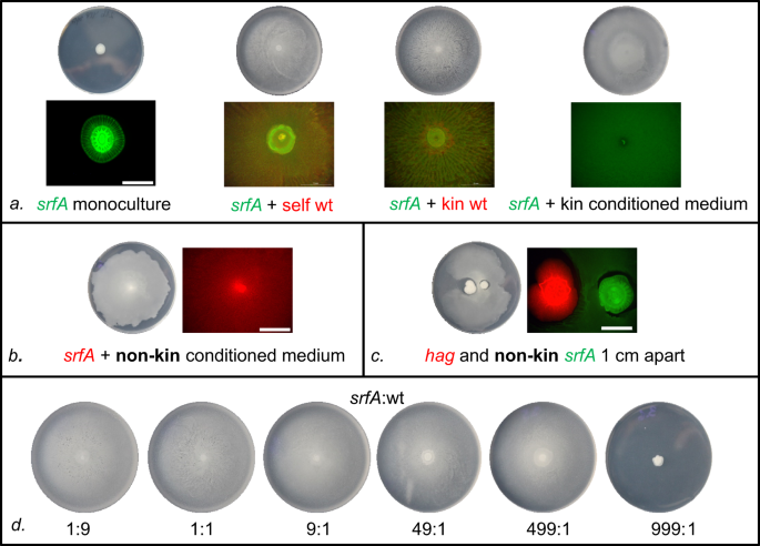
The srfA mutant with different types of strains that had been mixed with surfactin (PS-216) was conducted, with kin or nonkin in swarming agar. In part a, it contained the srfA by itself, mixed with PS-216 mKate which is the self combination, with PS-68 being the kin and lastly the conditioned medium contained 10 microliters from PS-216 mKate. In b the mutant contained the nonkin in a contained medium. In c hag and nonkin, srfA had a distance of 1cm, d is the swarming proportions at low proportion of wildtype.  Figure taken from Kraigh et al. 2021. 

Microbes are an essential part of life. They are of great importance because they not only shape microbial communities but also biofilm or swarms. The role was to investigate whether B. subtilis soil happens to have kin discrimination in their community. Different types of strains were used, the focal strain which was PS-216, and its mix partners (PS-68 kin wildtype), along with srfA mutant were inoculated on swarming agar. The results showed that in the absence of srfA, PS-216 didn't seem to swarm, but when the strain (kin) was introduced it swarmed over the entire plate. In the figure, A contained srfA monoculture, srfA, and the self wildtype, srfA and kin wild-type and srfA with kin continued medium. The kin with the srfA as discussed worked best. In part, the srfA with nonkin conditioned medium was spread across the plate similar to kin since both are relatively similar and concluded that in order for the kin to discriminate against the nonkin a direct contact between both cells has to happen. They then tried to prove this by proceeding with C concluding that even in absence of the cell to cell contact the nonkin can share surfactant due to the hag mutant.  D on the other hand concluded minor surfactant is needed for the populations to gain cooperative binding. Although surfactin did grow on the nonkin strain and was able to restore swarming of its surfactant, the territorial exclusion between that of nonkin as effective exploitation of public good. It was concluded that territorial exclusion only occurs among nonkin but not between kin. Overall more research should be conducted on whether the different types of methods using kin discrimination yield the same results, how can kin discrimination come to benefit and harm microbial activity as well. 

Original Article:
Kraigher, B., Butolen, M., Stefanic, P., & Mulec, M. I. (2021). Kin discrimination drives territorial exclusion during Bacillus subtilis swarming and restrains exploitation of surfactin. ISME J. https://doi.org/10.1038/s41396-021-01124-4


Water Quality Adversely Affects Poultry


Bacterial abundance of water samples in four series: V1, V3, V5, and V7. Data divided between (a) phyla and

(b) genera.  Figure taken from Wan et. al 2021. 


Water is critical to sustain our life on earth and its quality is an important factor to maintain a healthy ecosystem. As our planet observes a steady increment in the population, various industries have to modernize the way they function to provide their products in an efficient manner. This modernized change can be denoted in water systems, many commercial laying farms have adapted from open water systems to V-trough water systems with the purpose of decreasing labor, water leakage, and microbial contamination. Although water quality is regulated, commercial farms have overlooked the sanitation of the V-trough in water systems. The study conducted by Wan and his colleagues, analyzed the water of V-trough and tested for microbial abundance and activity in commercial laying farms by the usage of a quantitative real-time polymerase chain reaction (qRT-PCR) and 16s rRNA sequencing technology. Water samples were obtained in 24 distinct sections within four tiers: 1st, 3rd, 5th, and 7th. These tiers demonstrated a significantly high microbial abundance in V-trough rather than a water pipe. Subsequently, the phyla level demonstrated a high abundance in Proteobacteria and in the genera level the total abundance exceeded thirty percent in: Acinetobacter, Streptococcus, Rothia, and Comamonas. Many of these bacteria are sources of diseases that can decline the health of poultry and put at risk one of our major food sources. The findings of this study provide light to ensure the water quality of poultry is not neglected and encourages different techniques to control the contamination in water. Poultry amongst other animals provide daily aliments for many of us to survive, the least that we can do is ensure that our poor choices do not adversely affect them. 


Original Article:


Wan,Y., Ma, R., Chai, L. et al. Determination of bacterial abundance and communities in the nipple drinking system of cascading cage layer houses. Sci Rep 11, 19169 (2021).

https://doi.org/10.1038/s41598-021-98330-z


Urease positive bacteria caused by Breast Milk Urea

 By: Karla Hinojosa 

Figure. A) Specifies the type of urease gene clusters that are found in B. infantis. B) A software application was used to generate a phylogenetic tree that showed the urease subunit alpha genes that were expected to be located in the human stomach. C) Metagenomic urease gene hits that were retrieved from urease protein subunits. D) Ratio of reads showing the urease proteins of all the bacterial species depending on infants being breastfed or through formula. 

In infants, human milk supports the growth of a beneficial microbiome. Human milk contains urea. The study investigates whether the microbiota can utilize urea from human milk, as well as how the microbiota restores and uses nitrogen from breast milk. It is critical since infants rely on feeding and are required for health reasons. The microbiome assists infants in receiving amino acids and vitamins, as well as gut maturation and immunological development. In order to feed bacterial demands, bacterial urease can play a role in the gut nitrogen cycle. Nitrogen, particularly urea, has a significant impact on the human gut flora. In comparison to formula-fed infants, breast-fed newborns had a higher urea level. Urease-related genes were studied using an infant gut metagenome database. Urease-related hits, such as urease protein subunits, urea transporters, urease accessory proteins, and urease activity regulating genes, were filtered out of the alignment findings. Bifidobacterium longum subsp. Infantis and Bifidobacterium breve were cultivated on a special Bifidobacterium agar medium designed for counting Bifidobacterium species. A colorimetric urea assay was used to determine the urea levels. Five Bifidobacterium urease genes were discovered in seven species related to the human gut, one of which is Bifidobacterium infantis. The metagenome revealed a total of 27 taxa for urease protein components, with Bifidobacterium accounting for 9% of the total. Further research concluded that B. infantis may utilize urea as its primary nitrogen source. Urease can be used as a growth factor to use urea as the main nitrogen source. Urea in breast milk can shape a microbial potentially beneficial strain is based on the discovery that urea in breast milk can influence a microbial population. As a result, Bifidobacterium urease is an important component of the microbiota that uses urea in breastfed newborns. Bifidobacteria is essential for the well-being of infants. 

Original Article: 
Schimmel P., Kleinjans L., Bongers S. R., Knol J., Belzer C. (2021). Breast milk urea as a nitrogen source for urease positive Bifidobacterium infantis. FEMS Microbiology Ecology, Volume 97, Issue 3, fiab019, https://doi.org/10.1093/femsec/fiab019


Microbial Communities In Permafrost: Environmental Controls and Impacts

 By: Andrea R. Ortiz 


The figure above is from Alekseev et. al., 2020. Soil microbial communities at phylum taxonomic level within collected soil samples. 


    In Antarctica only about 0.1% of the region is considered ice free due to extreme temperatures and environmental conditions.  One ice free portion is found in Eastern Antarctica known as the Larsemann Hills. The permafrost of the Larsemann Hills is home to various microbial communities which are adapted to live in such extreme conditions. In a recent investigation of  Alekseev et. al., 2020 , various microbial communities were identified in the region along with their relationship to soil and impacts on the surrounding environment. 
  Using 3 sampling points of 50 x 50 m, a collection of soil samples from these sites were collected at various depths at dimensions of 20 x 20 cm. Each sample was taken in a double sterile bag to a laboratory where the soil was sieved before undergoing chemical analysis for pH, carbon - nitrogen content, and particle size distribution. The microbes identified in these conditions were found in a neutral pH due to the lack of salt accumulation, 10.48 and 16.33 carbon - nitrogen ratio because of peat material found in topsoil, and a distribution in sub-surface horizon due to fine earth formation. The individual microbes after analysis then underwent 16S rRNA gene pyrosequencing resulting in 12 bacterial and archaeal phyla being identified. Some of the collected samples included microbes such as Proteobacteria, Actinobacteria, Acidobacteria, Cyanobacteria, Gemmatimonadetes, Verrucomicrobia and many more. 
   The results of the study indicate that diverse soil microbial communities not only exists in such harsh conditions but are imperative to sustaining their ecosystem which include cycling of nitrogen,  determining carbon availability, and soil composition at multiple levels. However, in the study it was also observed that microbes that survive in extreme cold are ultimately impacted by moisture content in their environment and nutrient availability. Thus, future studies such as this should continue in order to comprehend microbial communities and effectively sustain the Antarctic ecosystem.   


Reference: 

Alekseev, I., Zverev, A., & Abakumov, E. (2020). Microbial communities in permafrost soils of Larsemann Hills, eastern Antarctica: environmental controls and effect of human impact. Microorganisms, 8(8), 1202.


C. elegans to-go microbes from slugs

 By. Sangwon Sloman-Moll


The figure shows the persistency of microbes through slugs to c. elegans; MYb115 is shown with green color from slug to c. elegans, and MYb 11 is shown with red color from slug to c. elegans. The color represent the presence of the microbes in the organism. Figure taken from Pees el al. 2021.

C. elegans is one of common model organism that used by researchers for human genome, and it is functional counterparts in humans which makes it an extremely useful model for human diseases. Microbes are also close related with human diseases. The paper by Pees and colleagues shows microbes can be persisted through organism to another organism. C. elegans use slug as a vector. However, C. elegans also acquire microbes from the vector. Pee and collagues compared C. elegans gut microbiomes isolated from wild-caught slugs to the microbiomes of worms after experimental slug passage to compare similarities and differences in microbiome composition. They found that microbiota persists in C. elegans while passing the slug gut and that worms simultaneously acquire additional bacteria species from the slug which shows on the figure. Therefore, the result demonstrates that C. elegans can take advantage of its passage through the slug by acquiring new potential microbiota without losing its native microbiota. The research about microbes on C. elegans recently started, yet studying the microbes on the C. elegans can advance the knowledge of microbes that persistence through different organisms. The future research on the subject would be the key to find the solution for parasite disease caused by the microbes.

Original article:

Pees, B., Johnke, J., Möhl, M., Hamerich, I.K., Leippe, M. and Petersen, C. (2021), Microbes to-go: slugs as source for Caenorhabditis elegans microbiota acquisition. Environ Microbiol. https://doi.org/10.1111/1462-2920.15730